In the earliest stages of development the embryo consists of a ball of cells, within which three layers can be easily identified.
- The outer layer, Ectoderm, will develop into the skin and central nervous system.
- The innermost layer, Endoderm, will form the lining of the gastro-intestinal tract.
- Between these is the Mesoderm, which will form the muscles, bones and many of the organs of the body.
The earliest sign of the nervous system is the development of the neural plate on the dorsal surface of the ectoderm, behind a column of cells known as the Notochord. The notochord is present in all vertebrates and is essential for inducing the development of the neural plate.
At the lateral edges of the neural plate, ridges appear that grow and fold towards each other to form a tube, the neural tube. The cells that lead this development are called Neural crest cells (C), and when they have completed their role in forming the neural tube, they go on to form some more specialised parts of the nervous system including the dorsal root ganglia, the autonomic nervous system and the adrenal medulla (D).
Thus the Neural tube is formed from the primitive Ectoderm of the embryo, and its formation depends on the presence of the notochord; the notochord is also important in the development of primitive nerve cells in the ventral half of the neural tube into motoneurones. The neural crest is important in ensuring the closure of the neural tube. This process starts in the middle section of the embryo, and the closure of the neural tube progresses towards the head and tail ends simultaneously. In humans this process is normally completed when the embryo is about 28 days old. |
Diagram of the Neural Plate and the formation of the Neural Tube.
|
|
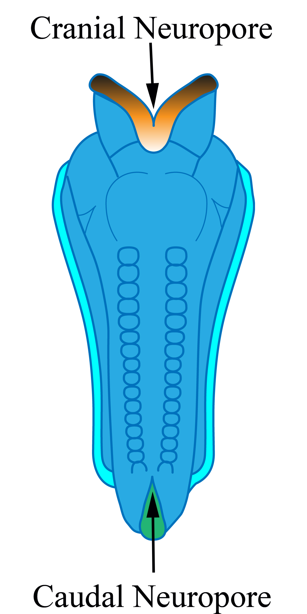
As the neural tube is forming, the central region closes first, and for a short time, the neural tube is open both at both ends, and the openings are called neuropores, these close completely around 28 days in the human.
The diagram shows the cranial and caudal neuropores before closure of the neural tube. Once closed, the neural tube becomes segmented, and each segment is called a somite.
After closure, the cranial end of the neural tube develops into primitive brain which can be divided into four distinct regions:
the prosencephalon,
which becomes the forebrain
the mesencephalon,
which becomes the midbrain
the rhombencephalon, which becomes the hindbrain and cerebellum, in addition tothe spinal cord.
Top
Neural Tube Defects arise of the failure of closure of the neural tube are not uncommon and present long lasting problems for the individual, if they survive. Failure of the neural tube to close at the front end results in anencephaly, and at the lower end, spina bifida.
The neural crest cells of the embryo migrate to other areas of the body and become specialised cells in the adult, forming for example the autonomic nervous system, the dorsal root ganglia and the adrenal medulla.
In other parts of the adult nervous system this central canal forms the ventricular system and both contain cerebrospinal fluid.
The spinal cord is a segmented structure, but the parts of the neural tube above the spinal cord, i.e. the brainstem, develop large expansions of nervous tissue that have specialised functions.
|
|
| Top
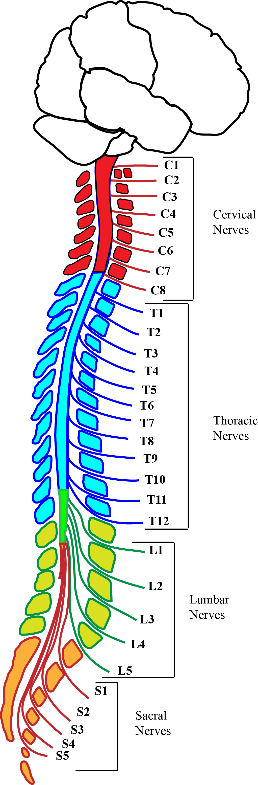
The spinal cord is segmented with 8 cervical, 12 thoracic, 5 lumbar and 5 sacral segments in humans. When the caudal neuropore fails to close, the deficiency is usually in the lumbo-sacral segments of the spinal cord.
The cranial end of the neural tube develops into massive structures in the adult human:
- the prosencephalon,
which expands enormously to become the cerebral hemispheres, basal ganglia,
thalamus and
hypothalamus
- the brainstem, consisting of the mesencephalon (midbrain) and the rhombencephalon, which becomes the hindbrain to which the cerebellum is attached.
|
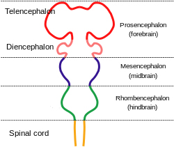 Diagram of the Embryological Origins of the Central Nervous System. |
| Top
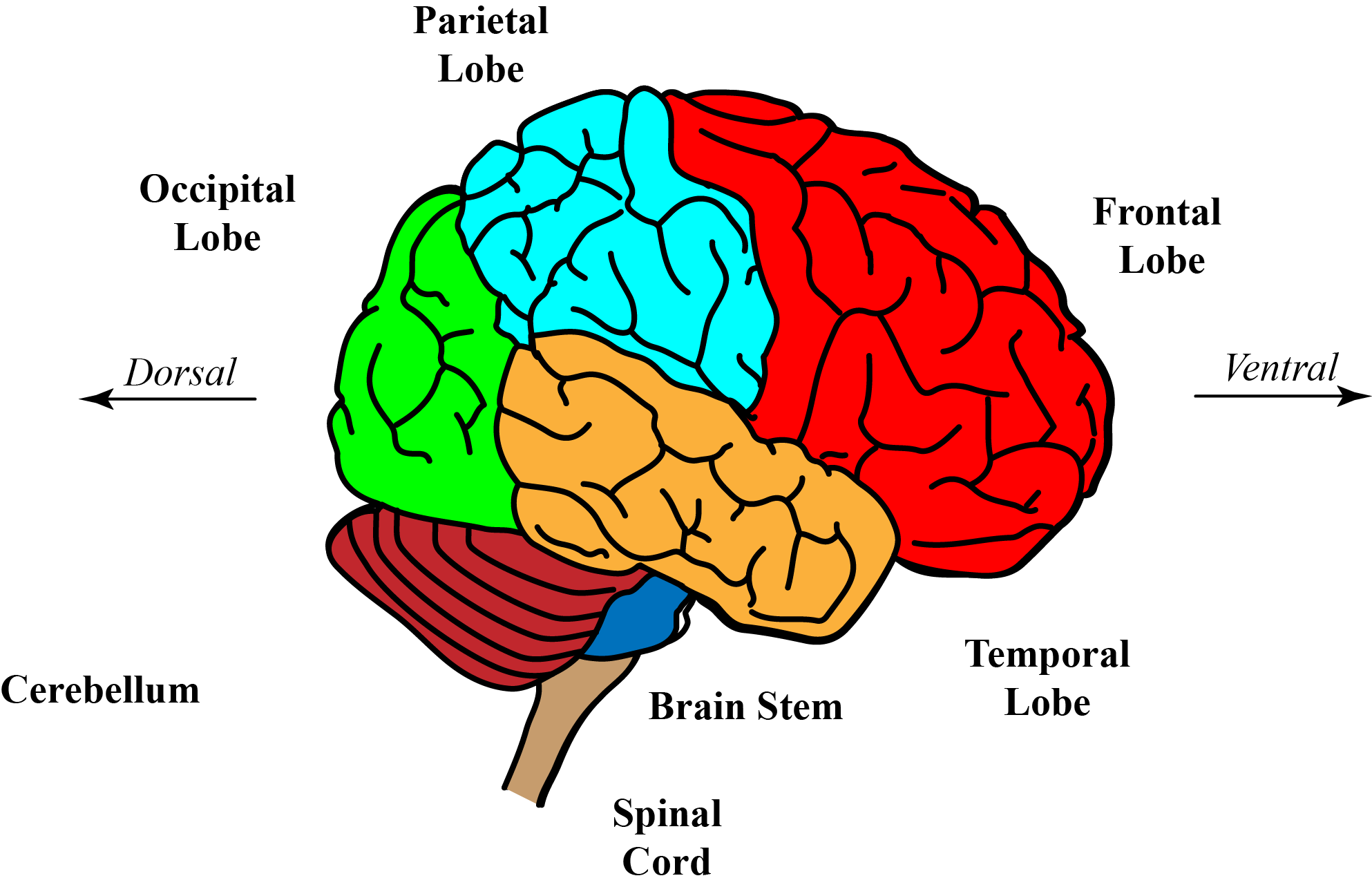
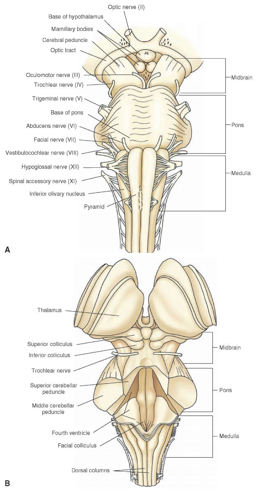
The brainstem is divided into three levels, the medulla (immediately above the spinal cord), pons and midbrain, through which nerve cells pass from the cerebral hemispheres to the spinal cord.
The medulla and pons of the embryo are sometimes called the hindbrain or rhombencephalon; just as the midbrain is sometimes called the mesencephalon.
The brainstem can be likened to a stalk that has some specialised appendages attached to it. One such large appendage, the cerebellum, bridges over the back of the pons, connected to it by a cerebellar peduncle on each side.
The cerebellar peduncles are composed of masses of nerve fibres that allow communication between the cerebellum with the spinal cord and the rest of the brain. Here the central canal is expanded as the fourth ventricle.
At the top of the brainstem, the ventral side of the midbrain consists of the cerebral peduncles, one on each side; these are the stalks which support and connect lower levels of the nervous system with the forebrain - the cerebral hemispheres and their central nuclei.
The cerebral peduncles (crura) contain nerve fibres that connect lower levels of the nervous system with the large nuclei in the centre of each cerebral hemisphere (the thalamus and basal ganglia) and the cerebral cortex. Each cerebral peduncle also contains a dark pigmented band of neurones - the substantia nigra, which is of importance in the control of voluntary movement.
On the dorsal surface the midbrain, behind the aqueduct (the analogue of central canal) exist four colliculi (small bumps- literally 'small hills'- on the surface of the tissue), two on each side, with specialised functions related to eye movements and hearing.
The two appendages that develop at the level of the thalamus are the optic cups that develop into the retinas and optic nerves of each eye. The Optic Nerve connects the retina and a specialised area of the thalamus - the lateral geniculate body or nucleus - which is easily visible and concerned with the processing of the visual image.
The thalamus and the area immediately below it, called the hypothalamus, are sometimes referred to as the diencephalon. The Optic Chiasma is an X-shaped structure where some optic nerve fibres from the retinas cross over to the opposite side of the brain, and can be seen clearly at the front end of the hypothalamus in the upper diagram opposite.
The forebrain or telencephalon consists of bilateral expansions at the front of the midbrain, and form the cerebral hemispheres and thalamus of the adult.
|
 * *
|
|
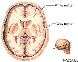
To the naked eye the brain and spinal cord appear a white and pinkish grey gelatinous mass, but when opened with a knife, there is a clear separation between two types of matter- grey and white.
The white matter consists of the myelinated axons of neurones, whereas the grey matter consists largely of neuronal cell boides and axonal terminals.
In the cerebral hemispheres the gray matter is on the outside of the brain and the white matter is inside.
This arrangement is reversed within the spinal cord, whose surface is white, consisting of axons passing along its length and connecting it to the brain.
|
 * *
|
 *
*